Always seek Physician consultation for any medical illness or problem.
Only a physician or licensed medical provider can diagnose medical conditions.
Please see the instruction section on the proper technique when using an otoscope for the exam of the ear canal: Please read carefully.
INSTRUCTIONS
Doubtless, you will find it useful to have an otoscope available in your home for examination of the ear canal. The best way to practice using your otoscope is with the help of an adult volunteer who has ear canals relatively free of ceruman (ear wax). To see the eardrum, grasp the outer ear. Gently pull backward and slightly upward on the ear (see ear picture below).
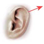
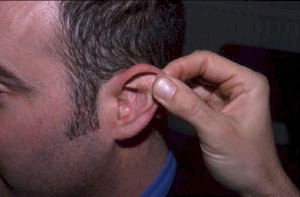
This will help to straighten the ear canal for the best "line of sight." Gently insert the otoscope while looking into its lens. You will begin to see when structures inside the ear come into focus. The focal length for optimal visualization of the eardrum varies upon the size of the ear canal. The length of the ear canal is variable in each and every person so it is important to watch closely through the lens while inserting the specula tip into the ear canal. This way you will know the instant your focal length is ideal and thus focus on the eardrum is at it’s best. (Never pry or force the otoscope into the ear canal).
Most physicians and nurses will tell you the way they learned to do otoscope exams was by practicing on each other back in medical and nursing school. The same holds true in this case. Find some willing adults and start looking into ear canals. Soon you will recognize what is usual and normal appearing. After you mastered the adult exam then you can move on to children and infants. Be patient in the learning process. Included you will also find 3 sizes of specula. It is recommended to begin your exam in adults with the largest diameter specula and move downward in size if needed. In children begin with the smallest diameter specula and move up in size if able.
You should be aware that it is occasionally impossible to see clearly into the ear of an infant, small child, or even a rare adult, with the most expensive professional otoscope. This is because the canal cannot be straightened sufficiently or it is occluded with ceruman (ear wax) or debris.
Usually, the view that is attainable is a function of ear canal size, and the presence of ceruman build-up. As a physician of many years experience, I assure you that virtually anything can be seen with our otoscopes that can be seen with the expensive wall mounted Welch Allyn otoscopes that I use at work.
The wall mounted Welch Allyn’s I use in the ER use 110 volt electricity as their power source. The portable models we sell use batteries, it is important to use fresh, strong batteries to maximize your light.
IMPORTANT: If you normally where glasses or contacts please leave them on while looking through your otoscope. We have had customers contact us saying that they could not see clearly because the instrument focused to close to the end of the specula. 98% of the time these were customers who were nearsighted and they had removed their glasses before looking into the lens of the otoscope. Occasionally we have had an issue with a defective lens. It is also important to not hold the otoscope too far away from your eye when looking into it.
If you are having any problem focusing I will include a quick and easy way to test to see if the otoscope lens is functioning properly or if the problem is simply user error or an ear canal to difficult to see in to due to earwax, debris, or curvature.
Basically an otoscope is a magnifier and a light whose job it is to focus clearly at approximately 1/2 to 3/4 inch from the tip of the specula. The test below allows you to tell if the otoscope is doing its job correctly.
Using the otoscope to look at newsprint you can quickly tell if there is a possible lens defect causing things to not focus properly. The eardrum is normally around 1 inch away from the entrance to the outer ear in adults and ¾ inch away in small children. When you subtract out the ¼ inch you normally insert the otoscope speculum into the ear canal your focal point should be between ½ and ¾ inches away when you look at the eardrum with the otoscope. You can test if the otoscope is functioning properly by viewing print from a magazine or newspaper and measuring the approximate distance the otoscope tip is away from the print when everything is in clear focus. It should be somewhere between ½-3/4 inch away. The print should also be very clear and not distorted. Please let another member of your family complete the test as well and see if they get a similar result as your test. Please notify us immediately if it appears your otoscope has a lens defect or any other problem.
BOTTOM LINE: If the light is bright and the focus on the newsprint is clear at 1/2-3/4 inch from the tip of the specula then the reason you are not seeing the eardrum clearly while doing an exam is not the fault of the otoscope. There are other factors causing this (i.e. earwax, debris, or ear canal curvature). If the newsprint is blurry or the light dim then there is a defect in the otoscope.
Sincerely,Doc Jim
FREQUENT QUESTIONS
Is it difficult for parents to learn to do ear exams?
This is one of those questions that has two answers....yes and no. The key to doing ear exams is practice. The old saying practice makes perfect could not apply more to any situation than it does to doing ear exams. It is important to begin doing otoscope exams on a willing adult as opposed to a child. The ear canals are larger and the eardrum is easier to see in an adult. The key is to look into as many adult ear canals as possible to get a feel for what is normal. Then when you visualize something that is not normal you can seek the help of your Doctor to advise you and diagnose a medical condition if one exists. Please remember it is your Doctors job to make a diagnosis. They spend many years in school and often many years in practice learning their profession.
Always go slow and never ever force or pry the otoscope in an ear canal in any way shape or form. Always look to see what is in front of you through the viewing window of the otoscope before advancing it into an ear canal. Never push the specula tip into the ear canal unless you have a clear view that there is nothing in front of you. It is also a good idea to have someone stabilize the head of a small child or infant since they can and will often jerk their head or pull away when something strange is being inserted into their ear. Again much of this is good old common sense, always remember to go slow and never push or pry with the otoscope.
Realize that in some children and even adults it is impossible to see the eardrum. Some children and even adults have very small ear canals and/or also filled with earwax and debris which make it impossible to see the eardrum. Even as a physician it is impossible to see into some ears.
It is also advisable especially with pediatric exams to get the help of your local pediatrician. Many pediatricians today are very supportive of home ear exams and recognize the value of parents being able to exam their children's ear canals with an otoscope. Pediatricians also recognize the importance of the early recognition of earwax occlusions that can cause hearing loss. If not recognized early this hearing loss can go on to affect the speech development in young children.
Will Disposable Specula work with the Dr Mom Otoscope?
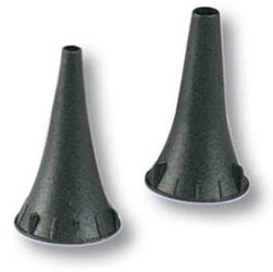
Yes our otoscope works well with disposable specula and are essential if you want to use the otoscope among multiple people We have disposables for sale for both the Dr Mom 4th gen and Dr Mom Pro as well as our smaller home use otoscopes.
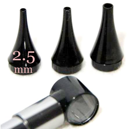
Disposable specula for our Third Generation Dr Mom LED and our Dr Mom Original Otoscopes
We have 3 size disposable specula available on our website at www.otoscopespecula.com and on Amazon
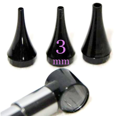
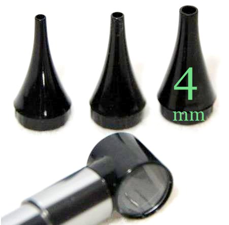
Disposable specula for our 4th Generation Dr Mom LED and our Dr Mom Pro Otoscope
We have a large 4.75mm Adult and a small 2.75mm Pediatric specula on our website at www.otoscopespecula.com and on Amazon
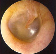
NORMAL EARDRUMS
Notice the different shades of color yet the eardrum still remains an opaque translucent appearance in all the pictures
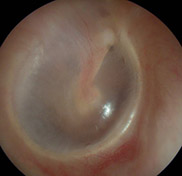
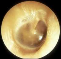
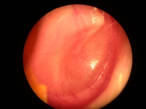
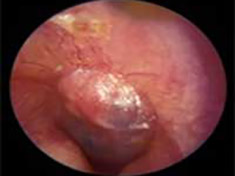
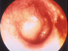
ACUTE INFECTION
Acute Infection with bulging of the tympanic membrane due to pressure from purulence (pus) behind it. The last picture reveals an ear tube that has gotten prematurely blocked and the ear is once again infected.
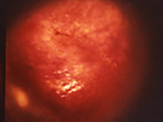
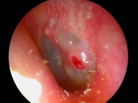
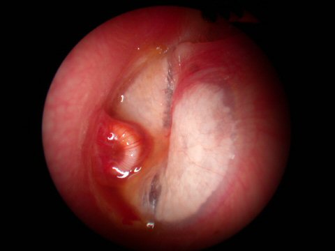
EXTERNAL OTITIS
External Otitis - Swimmers Ear This is an infection of the ear canal itself. Notice the swelling of the ear canal.
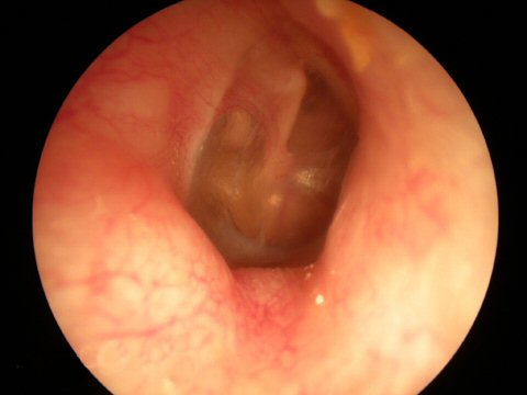
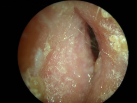
FUNGAL INFECTION
Fungal infection of the ear canal
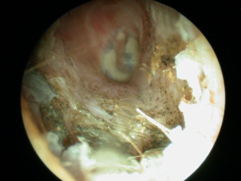
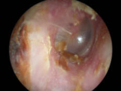
EAR TUBES
Ear tubes - The first picture shows an ear tube that has fallen out but is stuck to ear canal by ear wax. The last picture shows a T-tube, these stay in place much longer but run the risk of leaving a permanent perforation when finally removed.
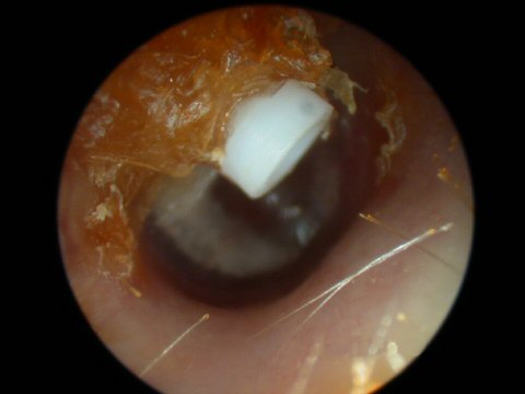
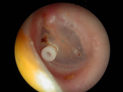
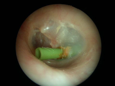
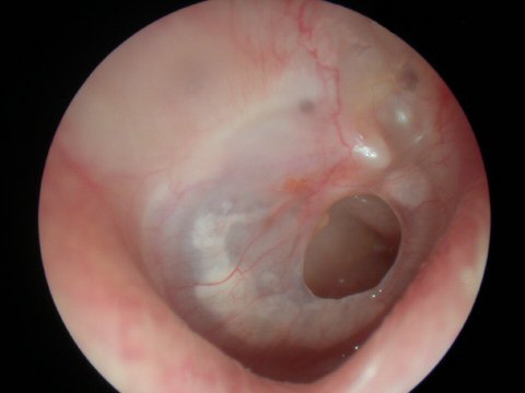
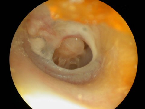
EAR DRUM PERFORATIONS
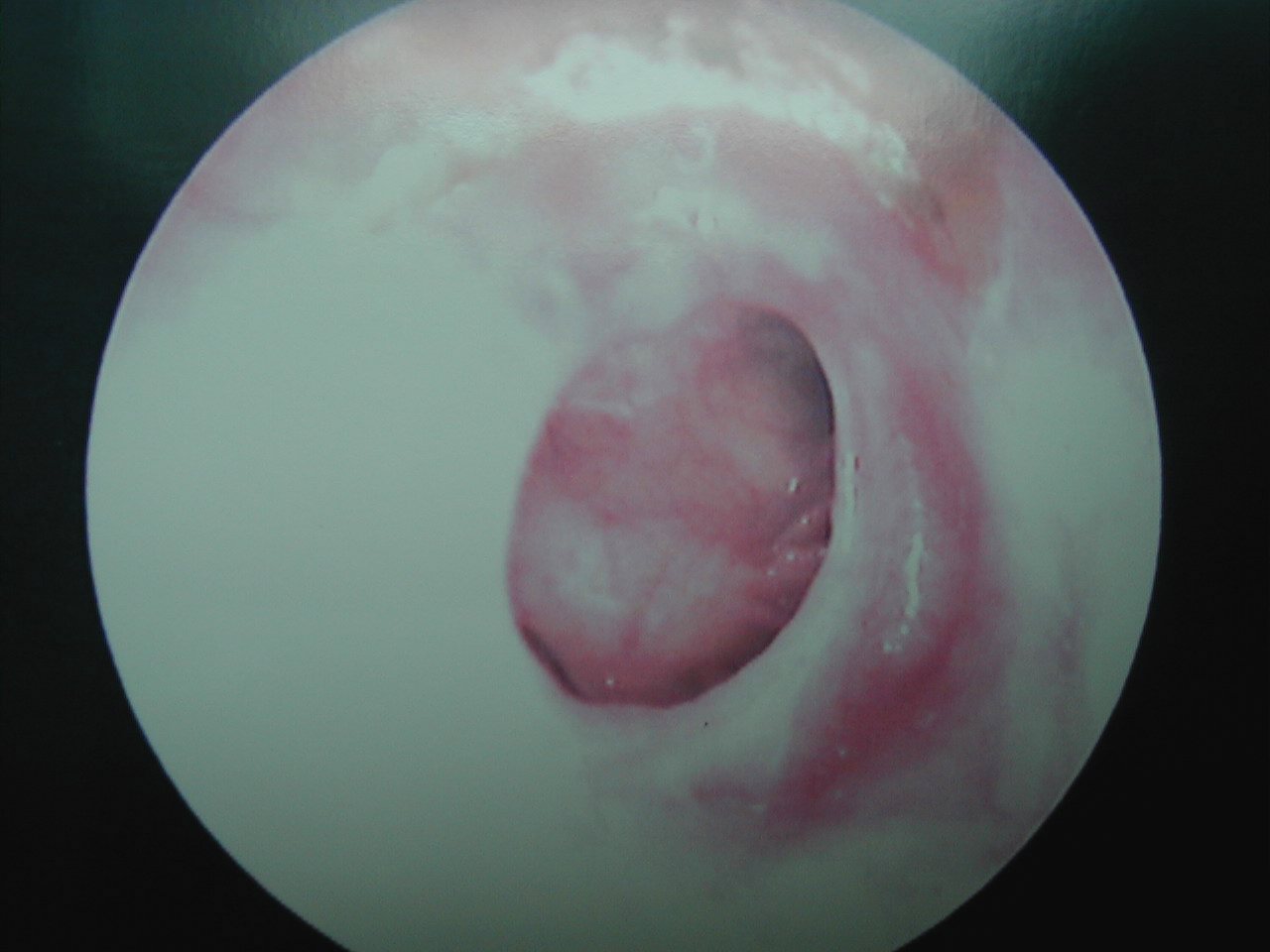
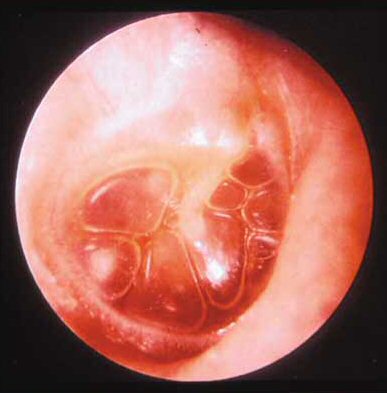
SEROUS OTITIS
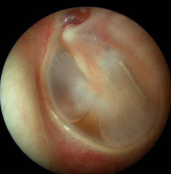
EAR WAX
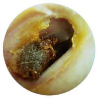
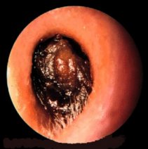
Ear Wax - notice the last picture shows an ear canal totally occluded with wax. This can be very harmful in a young child that is just learning to speak due negative affects hearing loss can have on speech development.
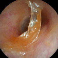
FOREIGN BODY
DISCLAIMER
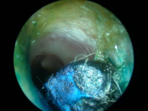
These images are for informational purposes only unless you are a physician or clinician who might use them as part of their medical training. Please remember only your Doctor can make a diagnosis of a medical problem. It is important to ALWAYS SEEK MEDICAL ATTENTION for any MEDICAL CONDITION. We have purposely shown more than one picture representing the same condition. You will find in the real world of eardrums that no two look exactly alike. This is why it is important to practice doing ear exams with your otoscope on as many willing volunteers as you can to get a feel for what is usual and normal. Also please realize these photos were taken with a $3,000 high resolution video otoscope so what you see with a standard hand held otoscope during an exam of an ear canal can not be expected to be as clear.